
About Us
| << Back |
“It’s just a PVD.”
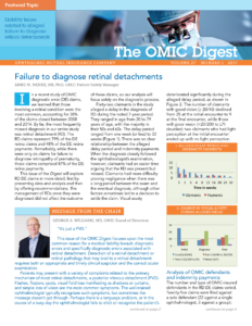
GEORGE A. WILLIAMS, MD, OMIC Board of Directors
This issue of the OMIC Digest focuses upon the most common reason for a medical liability lawsuit: diagnostic errors and specifically diagnostic errors associated with retinal detachment. Detection of a retinal detachment or retinal pathology that may lead to a retinal detachment requires both an appropriate and timely clinical suspicion and the correct ocular examination.
Patients may present with a variety of complaints related to the primary mechanism of most retinal detachments, a posterior vitreous detachment (PVD). Flashes, floaters, spots, visual field loss manifesting as shadows or curtains, and simple loss of vision are the most common symptoms. The well-trained ophthalmologist typically recognizes such symptoms, but sometimes the message doesn’t get through. Perhaps there is a language problem, or in the course of a busy day the ophthalmologist fails to elicit or recognize the patient’s symptoms. Patients may fail to appreciate monocular symptoms or visual loss through denial or neglect. Unrecognized or unmentioned trauma may not be described for social or personal reasons. Regardless, it is our responsibility to determine the patient’s problem.
Once a diagnosis of retinal tear or retinal detachment is suspected, the ophthalmologist must proceed with an appropriate examination. The question of what constitutes an appropriate examination is often the focus of a medical liability claim. The American Academy of Ophthalmology Preferred Practice Pattern (PPP) on Posterior Vitreous Detachment, Retinal Breaks and Lattice Degeneration (available at aao.org) is an authoritative, peer-reviewed summary of the standard of care. All ophthalmologists who see such patients should be familiar with these recommendations. I can assure you that any plaintiff’s lawyer will be.
A couple of common issues repeatedly arise. The first is the need for a dilated examination with binocular indirect ophthalmoscopy and scleral depression. Some ophthalmologists (and many patients) are uncomfortable with scleral depression. However, the PPP clearly states that scleral depression is the standard of care whenever a retinal tear or retinal detachment is suspected. The second issue involves a suboptimal view due to media opacification such as cataract, vitreous hemorrhage, miosis, or poor patient cooperation. In such situations, B scan ultrasonography is required. If the ophthalmologist fails to perform either test, there is cause for concern.
 Fortunately, the vast majority of people (myself included) with symptoms consistent with a retinal tear or retinal detachment will have an uncomplicated PVD. However, even after an appropriate examination confirms the absence of a retinal tear or detachment, the treatment process is not over. The ophthalmologist must instruct the patient and, importantly, the office staff concerning the need to return as soon as possible if there is a change in symptoms or vision. An office protocol concerning how to address such phone calls or patient contacts is a good idea and an example is available at www.omic.com. It’s hard to do the right thing if you don’t see the patient at the right time.
Diagnostic errors will always be inherent to the practice of medicine. That’s why you want a company with the financial strength and unsurpassed risk management programs of OMIC. The next time you think “It’s just a PVD,” remember one of Yogi Berra’s best aphorisms: “Never make the wrong mistake.”
Please refer to OMIC's Copyright and Disclaimer regarding the contents on this website


